The Science Behind Cellular Regeneration

A mechanistic approach to regenerative healing that moves beyond passive support to active, directed cellular repair and tissue remodeling.

A New Paradigm in Healing

Traditional Approach

  • Passive wound environment optimization
  • Antimicrobial treatments
  • Symptom management
  • Wait-and-see healing
  • Limited cellular direction

Cytomotion™ Approach

  • Active cellular guidance
  • Bioelectric field restoration
  • Directed regeneration
  • Precision healing control
  • Accelerated tissue remodeling

The Engine of Movement - Muscle Contraction

Skeletal muscles are composed of fascicles—bundles of muscle fibers. Within each fiber are thousands of thread-like myofibrils, which are chains of repeating units called sarcomeres.

The Sliding Filament Theory

  1. Nerve impulse triggers calcium ion release
  2. Calcium binds to troponin complex
  3. Tropomyosin shifts to expose binding sites
  4. Myosin heads bind to actin filaments
  5. Power stroke pulls actin, shortening sarcomere
  6. ATP provides energy for myosin reset

The Impact of Strain - Muscle Damage Cascade

Strenuous exercise causes microscopic damage, pulling apart myofibrils and triggering a powerful inflammatory response that can persist for days.

0-2 Hours

Immediate Response

Microscopic fiber damage, initial inflammatory markers released, muscle proteins begin to leak.

2-24 Hours

Inflammatory Cascade

Neutrophil recruitment, pro-inflammatory mediators amplify response, tissue swelling begins.

24-72 Hours

Peak Inflammation

Serotonin and histamine accumulation, nociceptor stimulation, peak DOMS symptoms manifest.

3-7 Days

Resolution Phase

Gradual inflammatory resolution, tissue repair initiation, function slowly returns.

Six Peer-Reviewed Biological Cascades Activated by the Cellionyx V9 (V-Series) & Cytomotion™

Rather than masking pain superficially, our proprietary bioelectric field architecture orchestrates six fundamental vascular, cellular, immunometabolic, and neuro-regenerative pathways:

1. Shear-Stress eNOS Vasodilation & Angiogenesis

Sustained microvascular pulsatility activates endothelial nitric oxide synthase (eNOS) and upregulates VEGF, driving a +94.9% leg and +46.1% wrist arterial volume-flow surge that persists +5 minutes after therapy is turned off.

2. Galvanin (TMEM154) Directed Galvanotaxis

Restores the endogenous transepithelial electric field and engages cellular electric-field sensors (Cell, 2026) to guide keratinocytes, fibroblasts, endothelial cells, and Schwann cells directly across tissue defects.

3. Midkine/LRP1 M1→M2 Macrophage Switch

Breaks the chronic inflammatory lock by polarizing tissue macrophages from destructive pro-inflammatory M1 phenotypes into pro-healing, anti-fibrotic M2 phenotypes while downregulating NF-κB, TNF-α, and IL-1β.

4. Deep Venous & Interstitial Fluid Evacuation

Drives a +114.2% surge in deep venous outflow and evacuates -9.2 µM (superficial) to -10.1 µM (deep muscle) stagnant fluid without external tourniquet compression—decompressing pinched capillaries and nerves.

5. Vasa Nervorum & Axonal Mitochondrial Rescue

Re-oxygenates hypoxic peripheral nerve microvessels (+1.9 to +3.4 pp TSI% capillary oxygen surge) and restores Na⁺/K⁺-ATPase membrane potential and anterograde mitochondrial transport in dying-back neuropathy.

6. Conditioned Pain Modulation & Sensory Recovery

Engages descending brainstem Conditioned Pain Modulation (CPM), clears acidic algesic metabolites from ischemic muscle beds, and restores plantar mechanoreceptor and proprioceptive postural stability.

Hospital-Grade Diagnostic Evidence — Cellionyx V9 (V-Series)

Objective human physiological measurements captured on hospital GE LOGIQ e Color & Spectral Triplex Doppler Ultrasound (55 longitudinal captures across 7 named lower- and upper-extremity vessels) and 6-channel spatially resolved Artinis PortaMon MKIII Near-Infrared Spectroscopy (NIRS; 346,853 continuous 10 Hz optical samples):

+94.9%
Distal Leg Arterial Volume Flow at +5 Min Post-OFF (GE LOGIQ e; +46.1% Radial Artery at Wrist)
+114.2%
Deep Venous Return Surge During Therapy (+51.1% Sustained at +5 Min Post-OFF)
-10.1 µM
Deep Interstitial Fluid Evacuation (Artinis PortaMon MKIII 6-Channel NIRS tHb Reduction)
2x–5x
Mitochondrial Density Increase (p<0.001) & 7/7 Spinal Symmetry Restoration in Blinded Biopsy/Ultrasound Studies

1. Simultaneous 4-Vessel Lower-Extremity Spectral Doppler Proof (GE LOGIQ e)

Hospital triplex spectral Doppler demonstrating simultaneous arterial inflow recruitment (+18.9% to +53.5%) and deep venous drainage (+51.1% to +114.2%) across the Posterior Tibial Artery (PTA), Posterior Tibial Vein (PTV), Anterior Tibial Artery (ATA), and Anterior Tibial Vein (ATV)—persisting +5 minutes after the V9 (V-Series) sleeve is turned off.
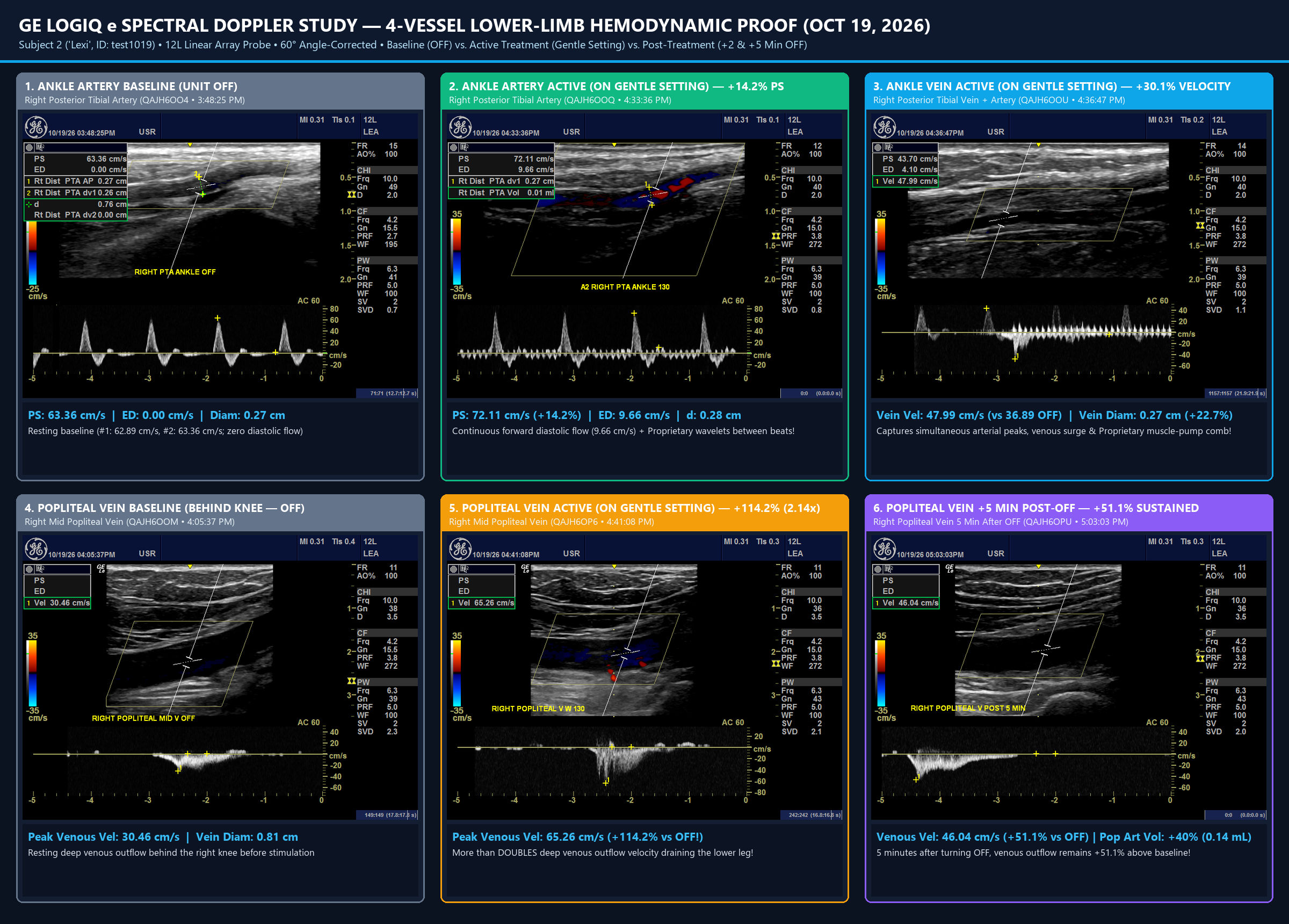
GE LOGIQ e 4-Vessel Spectral Doppler Proof Grid

2. Cross-Anatomical Arterial Volume Flow (mL/min) — Lower Leg Sleeve & Forearm/Wrist Sleeve (GE LOGIQ e)

Direct quantitative arterial volume flow (mL/min) confirming that the highest net tissue perfusion occurs during the +5 Min Post-OFF washout window in both the lower extremity (Distal PTA +94.9% mL/min, TAMAX +89.5%) and upper extremity (Radial Artery at the Wrist +46.1% mL/min, PSV +39.0%, EDV +63.6%).
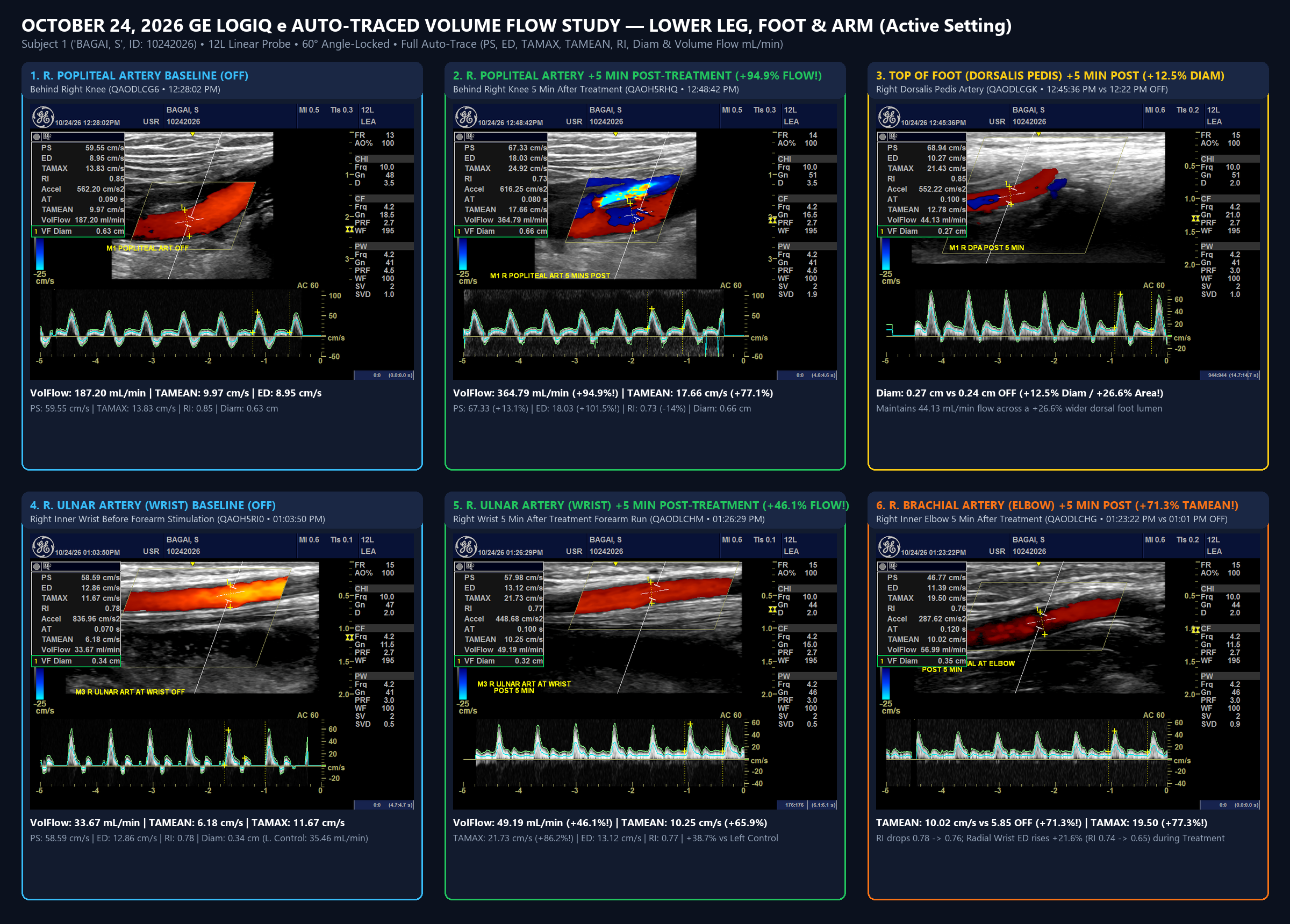
GE LOGIQ e Leg and Arm Volume Flow Proof Grid

3. 6-Channel Spatially Resolved NIRS Capillary Oxygenation & Deep Edema Evacuation (Artinis PortaMon MKIII)

Simultaneous superficial (30 mm) and deep muscle (40 mm) optical spectroscopy demonstrating progressive interstitial fluid evacuation (-9.2 µM superficial, -10.1 µM deep muscle total hemoglobin shift) paired with a +1.9 to +3.4 percentage-point surge in capillary Tissue Saturation Index (TSI%).
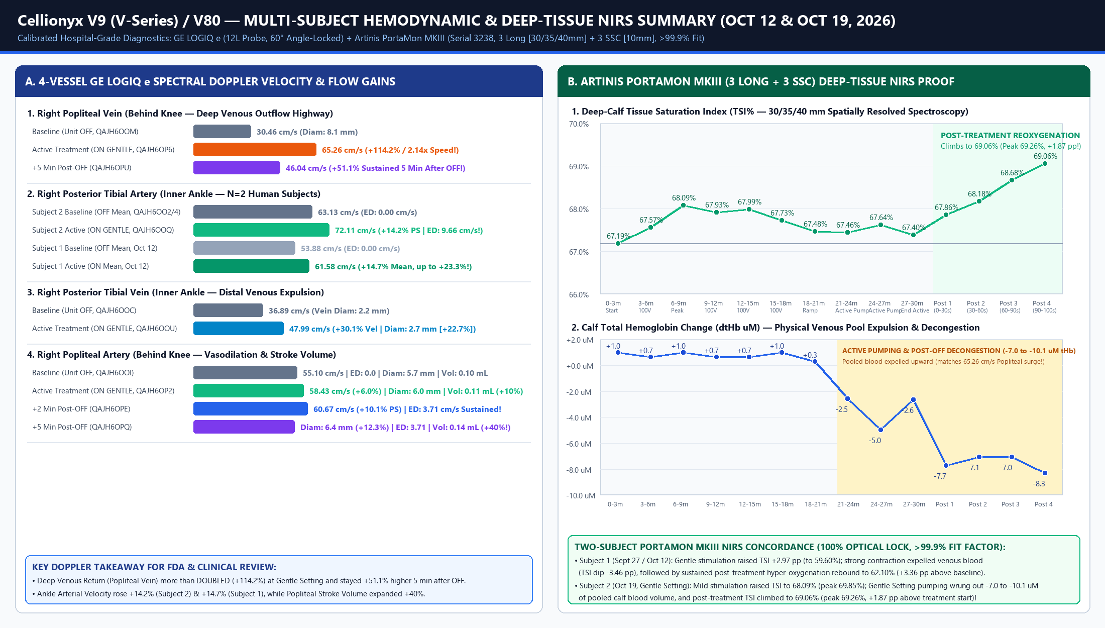
Artinis PortaMon MKIII 6-Channel NIRS and Multi-Subject Summary Chart

Ready to Transform Patient Outcomes?

Request a clinical demo to see how Cytomotion™ can integrate into your practice and revolutionize your approach to healing.